AP Biology DNA Replication: A Complete Guide for Students
Master AP Biology DNA replication with clear explanations of enzymes, leading and lagging strands, replication forks, and common exam misconceptions.

You're staring at a replication diagram, memorizing helicase, primase, polymerase, and ligase, but the arrows still don't seem to make sense. Then an AP Biology question asks why one strand is continuous, why the other requires Okazaki fragments, or how proofreading prevents mutations. Knowing the enzyme names isn't enough. You need to understand the logic that forces each step to happen.
DNA replication becomes much easier once you connect DNA's antiparallel structure, the 5′ to 3′ synthesis rule, and the need for accurate copying before cell division. This guide builds that reasoning from the ground up, then applies it to experimental evidence, enzyme functions, replication fidelity, and the kinds of explanations that earn points on AP Biology free-response questions.
Table of Contents
- What AP Biology Expects You to Know About DNA Replication
- The Discovery That Changed How We Think About Replication
- How Antiparallel Strands and the 5′ to 3′ Rule Shape the Fork
- Leading Strand Versus Lagging Strand
- The Core Enzymes You Must Know for the Exam
- Why the Lagging Strand Needs So Many Primers
- Replication Fidelity and What Happens When Repair Fails
What AP Biology Expects You to Know About DNA Replication
A replication diagram may show helicase, primase, polymerase, and ligase, yet an exam question can still ask what each enzyme accomplishes and why the steps occur in that order. The central purpose comes first: before cell division, a cell copies its DNA so genetic information reaches both daughter cells. Replication is semiconservative, so each new DNA molecule contains one parental strand and one newly synthesized strand, as explained in the OpenStax overview of DNA replication. Each parental strand provides the sequence information for building its complementary partner.
AP Biology expects you to connect that model to the sequence of events:
- Origins are recognized, marking where copying begins.
- Helicase separates the parental strands, forming replication forks.
- Primase makes RNA primers, which provide free 3′ ends.
- DNA polymerase extends new strands by adding complementary nucleotides.
- DNA ligase joins fragments after discontinuous synthesis.
- Proofreading and repair systems correct many errors that escape initial checking.
These steps are often tested through diagrams, strand labels, and predictions about the products of replication. The detailed reason that one strand is copied continuously while the other is assembled in fragments belongs to the fork analysis later in the guide. For the exam, first track the template, the new strand, the direction of synthesis, and the enzyme acting at each stage. The overall sequence is also summarized in OpenStax's AP Biology chapter summary.

What exam questions often test
You may need to identify a template strand, determine the direction of a new strand, interpret a replication diagram, or predict DNA products after successive rounds. Some questions also ask you to distinguish the conservative, semiconservative, and dispersive models. Keep the models separate: they describe different arrangements of parental and newly made DNA.
Strong FRQ responses explain cause and effect rather than listing enzyme names. State what the enzyme does, identify the strand or structure involved, and connect the step to accurate copying. Practice organizing these relationships with AP Biology flashcards.
Exam rule: Identify the strand, the template direction, the new-strand direction, and the responsible enzyme. A label alone may earn less credit than a concise explanation showing how those details fit together.
The Discovery That Changed How We Think About Replication

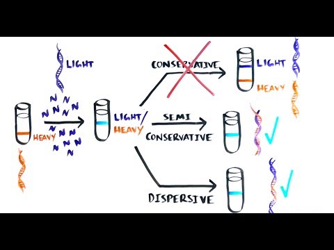
A DNA band appears in the middle of a centrifuge tube after bacteria switch from heavy nitrogen to light nitrogen. That result helped resolve a question scientists had debated: how are parental DNA strands distributed into daughter molecules?
Three models made different predictions. The conservative model kept the original double helix intact and produced a completely new double helix. The semiconservative model separated the parental strands, with each strand guiding synthesis of a new partner. The dispersive model mixed old and new DNA within both daughter molecules.
Matthew Meselson and Franklin Stahl tested these models in 1958 using 15N-labeled DNA and density-gradient centrifugation, as described in the historical review of the Meselson-Stahl experiment. They first grew bacteria in heavy nitrogen, which labeled existing DNA. They then transferred the bacteria to a light-nitrogen environment and examined DNA after successive generations.
Density-gradient centrifugation separates DNA by buoyant density. After one generation in light nitrogen, the DNA formed a single intermediate-density band. Each molecule therefore contained heavy parental material and newly synthesized light material. A conservative mechanism would have produced separate heavy and light molecules.
After two generations, the DNA formed intermediate-density and light-density bands. This pattern matched daughter molecules containing one old strand and one new strand. It supported semiconservative replication and excluded the conservative model, while further labeling analysis ruled out the dispersive model.
Why the evidence matters on the AP exam
The tested skill is reading what each band represents. If a question gives density bands after one or more generations, compare that pattern with the prediction made by each model before selecting an answer. The experiment addressed the arrangement of parental and newly synthesized DNA, not merely whether DNA can copy itself.
Arthur Kornberg supplied related evidence during 1956 to 1958. He isolated DNA polymerase and showed that the enzyme could synthesize DNA in vitro. His work established that replication is template-directed and enzymatic, rather than a direct duplication of one entire DNA molecule. Kornberg received the 1959 Nobel Prize in Physiology or Medicine for this work, according to the historical review of the Meselson-Stahl experiment.
Watch the experiment-focused explanation before practicing band-pattern questions:
How Antiparallel Strands and the 5′ to 3′ Rule Shape the Fork
The leading and lagging strands exist because DNA strands run in opposite directions. One parental strand has a 5′ end and a 3′ end, while its complementary partner runs from 3′ to 5′. This arrangement is called antiparallel orientation.
DNA polymerase adds a nucleotide only to a free 3′ hydroxyl group, so every new strand grows from 5′ toward 3′. The enzyme reads the template strand in the opposite direction, from 3′ toward 5′. This is the rule that controls everything at the replication fork.
As helicase opens the double helix, the two exposed templates point in opposite orientations. On one template, DNA polymerase can follow the moving fork while extending the new strand continuously. That product is the leading strand. On the other template, polymerase must work in short sections that extend away from the fork. That product is the lagging strand, as shown in the OpenStax explanation of replication forks.

Trace the ends, not just the arrows
Students often say, “The leading strand is 3′ to 5′.” That statement confuses the template with the newly synthesized strand. The template may be oriented 3′ to 5′ in the direction polymerase reads it, but the new leading strand is always synthesized 5′ to 3′.
Use this tracing routine when you see a diagram:
- Find the parental template.
- Mark its 5′ and 3′ ends.
- Determine where the new strand's free 3′ end is located.
- Draw synthesis only in the 5′ to 3′ direction.
- Ask whether the polymerase is moving toward or away from the fork.
The nitrogenous bases pair through complementary interactions, with purines pairing with pyrimidines. If that relationship needs review, revisit the purine and pyrimidine guide before returning to fork diagrams.
Practical rule: The direction of polymerase movement is not the same thing as the direction of DNA synthesis. Polymerase may move away from the fork on the lagging strand, but the strand it creates still grows 5′ to 3′.
Leading Strand Versus Lagging Strand
At one replication fork, the two strands are copied simultaneously, but they use different physical arrangements. The leading strand can be extended toward the fork as helicase continues opening the parental DNA. The lagging strand must be assembled behind the fork in pieces.
| Feature | Leading strand | Lagging strand |
|---|---|---|
| Synthesis pattern | Continuous | Discontinuous |
| Primer requirement | A primer begins synthesis | Each Okazaki fragment needs a primer |
| Polymerase direction | 5′ to 3′ | 5′ to 3′ |
| Relationship to fork | New DNA grows toward the fork | New DNA segments grow away from the fork |
| Final processing | Relatively direct extension | Primer removal, replacement, and ligation |
The leading strand
Once an RNA primer provides a starting 3′ end, the main replicative DNA polymerase can add nucleotides continuously as the fork advances. In many AP Biology diagrams, this strand appears as one long arrow pointing toward the fork. The arrow represents the direction of new-strand growth, not the direction of template reading.
The leading strand doesn't avoid the 5′ to 3′ rule. It benefits from the fact that its template orientation allows polymerase to keep extending the same growing end while helicase exposes more template.
The lagging strand
The lagging strand has the same polymerase and the same synthesis direction, but the template's orientation puts the growing end on the wrong side of the fork. Polymerase therefore makes one short segment, then another, and then another as additional template becomes available.
These segments are Okazaki fragments. Each fragment begins with an RNA primer, receives DNA nucleotides from polymerase, and is eventually connected to its neighbor by DNA ligase. The cell removes the RNA primers and fills those spaces with DNA before the remaining nicks are sealed.
A common wrong answer says that the lagging strand is made 3′ to 5′. It isn't. The lagging strand is synthesized 5′ to 3′ in separate sections. Another wrong answer claims that the leading strand is made by a different directional rule. Both new strands obey the same rule. Fork geometry creates the difference, not a difference in polymerase chemistry.
Point-saving explanation: “The lagging strand is discontinuous because DNA polymerase can extend only from a 3′ end and can synthesize only 5′ to 3′, while the parental strands are antiparallel.”
The Core Enzymes You Must Know for the Exam
A replication-fork diagram becomes easier when each enzyme answers a specific physical problem. Helicase opens the parental double helix. Primase lays down an RNA starting point. DNA polymerase III extends new DNA, while DNA polymerase I replaces RNA primers with DNA. DNA ligase seals remaining nicks, and topoisomerase reduces twisting ahead of the fork.
| Enzyme | Function | Substrate / Direction |
|---|---|---|
| Helicase | Separates the two parental DNA strands by disrupting hydrogen bonds between paired bases. | Double-stranded DNA at the replication fork |
| Primase | Synthesizes short RNA primers so DNA polymerase has a free 3′ end to extend. | DNA template; primer synthesis begins the new strand |
| DNA polymerase III | Adds complementary DNA nucleotides to the growing strand and performs the main replication work on both strands. | Template DNA and a free 3′ end; synthesis proceeds 5′ to 3′ |
| DNA polymerase I | Removes RNA primers and replaces them with DNA in the replicated molecule. | Primer-containing DNA regions |
| DNA ligase | Seals nicks in the sugar-phosphate backbone, joining neighboring Okazaki fragments. | Adjacent DNA fragments with a break between them |
| Topoisomerase | Relieves torsional strain and supercoiling that builds ahead of the replication fork. | Twisted DNA ahead of the fork; acts on the backbone |
Separate the commonly confused enzymes
Helicase and topoisomerase act in different locations. Helicase separates complementary strands at the fork by disrupting hydrogen bonds between paired bases. Topoisomerase works ahead of the fork, easing strain caused by the DNA's increasing twist. If a diagram shows tightly wound DNA in front of the fork, identify topoisomerase rather than helicase.
DNA polymerase III and DNA polymerase I also divide the work. Polymerase III carries out most DNA elongation on both new strands. Polymerase I removes RNA primers and fills those regions with DNA. Ligase performs neither task. It joins neighboring DNA after the correct sequence has been inserted, closing breaks in the sugar-phosphate backbone.
These roles also explain why the lagging strand requires repeated processing. Its short DNA sections each begin with a primer, and ligase must connect the completed sections. On an exam, connect the enzyme to the problem it solves rather than memorizing isolated names.
A useful study method is to make cause-and-effect cards. For example, ask, “If ligase is inhibited, what remains between Okazaki fragments?” The answer is a discontinuous sugar-phosphate backbone with unsealed nicks. Guidance on how to make flashcards online can help you turn enzyme functions into questions based on class diagrams.
Why the Lagging Strand Needs So Many Primers
DNA polymerase cannot begin a strand without an existing free 3′ hydroxyl group. Primase supplies that starting point by building a short RNA primer, which DNA polymerase can extend.
For AP Biology questions, the useful question is not “Why are there primers?” It is “Where must each primer appear?” On a diagram, locate the replication fork and mark the newly exposed lagging-strand template. Each exposed segment receives a primer near the fork. DNA polymerase then extends that primer to form an Okazaki fragment, while the earlier fragments remain behind the fork.
The number of primers therefore depends on how much DNA must be copied in separate fragments. A longer region requires more Okazaki fragments and more primers. If fragments are shorter, primer density increases. If fragments are longer, fewer primers are needed across the same region. Fork speed does not change the direction of synthesis, but it can change how quickly new template becomes available and how often primase must begin another fragment.
A common FRQ may show several primers and ask students to explain their placement. A complete response should connect three observations: primers appear repeatedly on the lagging strand, each primer starts one Okazaki fragment, and every fragment grows 5′ to 3′. Do not claim that the lagging strand is synthesized 3′ to 5′. That answer loses the point even if the diagram is labeled correctly.
After DNA polymerase fills the gaps, DNA polymerase I removes the RNA primers and replaces them with DNA. DNA ligase seals the remaining nicks between adjacent fragments, producing one continuous sugar-phosphate backbone.
Common misconception: Multiple primers do not indicate multiple synthesis directions. They indicate multiple starting points for fragments that all extend 5′ to 3′. This distinction helps you explain primer placement rather than merely identify it.
Replication Fidelity and What Happens When Repair Fails
A polymerase can copy a long DNA molecule quickly, yet one mismatched base can change a codon or disrupt gene expression if it remains. Cells therefore protect the sequence through several safeguards. Polymerase selectivity reduces mistakes to about 1 error per 10⁵ bases, and proofreading improves accuracy to around 1 error per 10⁷ bases, according to the AP Biology replication fidelity resource. After replication, mismatch repair lowers the final error rate to roughly 1 mistake per 10⁹ to 10¹⁰ bases per replication.
| Fidelity stage | Mechanism | Approximate error rate | Failure consequence |
|---|---|---|---|
| Polymerase selectivity | Correct base pairing and active-site selection | About 1 per 10⁵ bases | Incorrect nucleotides enter the new strand |
| Proofreading | Polymerase removes many mismatches after incorporation | Around 1 per 10⁷ bases | Some replication errors remain |
| Mismatch repair | Repair proteins detect and correct escaped mismatches | Roughly 1 per 10⁹ to 10¹⁰ bases per replication | Persistent mutations can contribute to genome instability |
Why several safeguards are necessary
Human DNA contains about 3 billion base pairs, so a rare mismatch can still matter when it escapes every correction layer. Proofreading checks the newly added nucleotide near the polymerase active site. Mismatch repair acts later, examining the newly replicated DNA and correcting errors that remain. These systems work like an inspection process with checkpoints: an early mistake can be caught immediately, and a missed mistake can receive another review.
For an FRQ, avoid writing that “DNA polymerase makes no mistakes.” State that polymerase occasionally adds an incorrect nucleotide, proofreading removes many mismatches, and repair proteins correct additional errors. That sequence earns more credit because it connects enzyme activity to mutation and inheritance.
When repair pathways fail
A replication error becomes biologically important when correction fails and the altered sequence persists. Its effect depends on its location and on whether it changes gene expression or the encoded protein. Weakened proofreading or mismatch repair can raise the mutation burden and threaten genome stability, linking replication to disease biology, aging, cancer, and other real-world consequences.
The AP Biology FRQ practice material on DNA, RNA, and protein synthesis reinforces the need to apply replication and repair concepts rather than list repair as an isolated vocabulary term. On an exam, describe the layered safeguards, then explain how repair failure allows a mutation to persist.
ClassLecture.ai can turn your own AP Biology lectures, notes, textbooks, or research papers into searchable transcripts, summaries, flashcards, and conversational Q&A about replication forks and proofreading. Upload your course material at ClassLecture.ai, then ask targeted questions about repeated primers on the lagging strand or practice explaining replication fidelity in an FRQ response.
The ClassLecture.ai Team
We build ClassLecture.ai, the AI study assistant that turns your recorded lectures into transcripts, summaries, flashcards, and answers cited to the exact timestamp — so you learn faster from your own professor's words.
Keep reading
Psych 101 Review: Core Topics, Practice Q&A, and Study Tools
A practical psych 101 review with core topics, key terms, sample Q&A, and study tools to help you prep for exams and master intro psychology.
ReadAP Microeconomics Unit 3 Study Guide with Practice Questions
Master AP Microeconomics Unit 3 with clear explanations of supply, demand, elasticity, and market structures, plus examples and practice questions.
ReadAP English Exam Study Guide: Read, Write, and Score Higher
A complete AP English exam study guide covering reading, writing, rubrics, practice prompts, and revision strategies to help you score higher in 2026.
Read